Email
EmailCase Study: Approach to Premature Closure of the Distal Radial Physis
Overview
Gayle Jaeger, DVM, MSpVM, DACVS, an orthopedic and soft tissue surgeon at VRC, has been treating a Golden Retriever puppy named Lito after his distal radial physis closed prematurely. As a result, his right radius was shorter than the adjacent growing ulna with subluxation of the elbow joint. If left untreated, this would progressively create lameness and complete dysfunction of the limb.
History & Diagnostics
Lito initially presented to Dr. Jaeger in early December 2016 at 5.5 months of age, with a progressive three-week history of right front limb lameness unresponsive to rest and anti-inflammatories. Radiographs at that time revealed evidence indicating premature closure of his distal radial physis with secondary elbow subluxation.
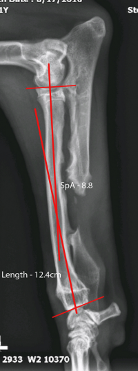
Initial Radiographs
 |
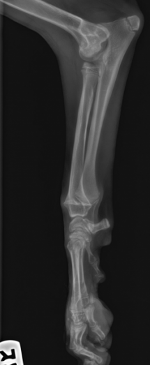 |
Above are lateral radiograph projections of the front limbs. The left is the normal side and the right is the affected side. Notice the open left distal radial physis on the left compared to the closed distal radial physis on the right. Also, note the shorter length of the right radius compared to the ulna as well as the resultant radiohumeral (elbow) incongruity.
Procedures
Partial Ulnar Ostectomy
A partial ulnar ostectomy was elected in an attempt to curb the secondary effects this incongruity would create on the elbow and carpal joints. By performing this procedure, we realized that over time his right antebrachium and overall limb length would be shorter than his left side. Our first priority was to save his elbow joint from discomfort and irreversible damage as the ulna continued to push up against the humerus.
 |
 |
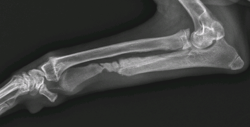
Above are post-operative lateral radiographs of the right antebrachium before and after the partial ulnar ostectomy. There is immediate mild improvement of the step between the height of the radius and ulna simply by releasing tension in the longer ulna. This improved further with weight bearing on the limb.
Second Partial Ulnar Ostectomy
Unfortunately, due to his young age, Lito’s ulna healed prematurely requiring a second partial ulnar ostectomy to remove more of the ulna while he continued to grow.
 |
Note above how the step between the radius and ulna improved with weight bearing, making his elbow more comfortable and preventing deformity.
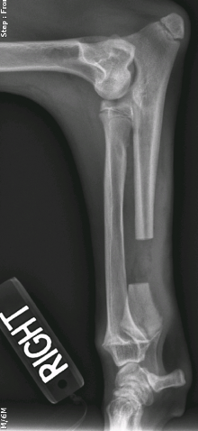
Once his growth plates started closing and we knew how long his normal leg would be as an adult, we could measure the true length deficit of his right side and start planning to lengthen his limb. The affected right radius was ultimately almost 4 cm shorter in length than the normal left side.
 |
 |
Above depicts the left fully-grown normal limb versus the shorter right limb. Note the persistent changes of his right elbow compared to his left.
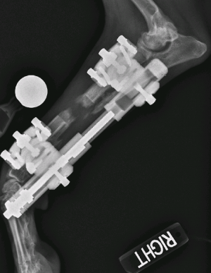
Radial Osteotomy & Stryker Triax External Skeletal Fixator Application
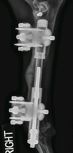
We contacted Stryker for assistance in providing a special external skeletal fixator called the Triax. This device would allow us to slowly lengthen his leg over time. The external connecting bar of the Triax frame has the ability to lengthen bone by turning a distraction bolt; separating two bone segments in small increments. Lito again went into surgery and we repeated the partial ulnar ostectomy, created a radial osteotomy, and applied the external fixator.
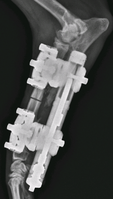 |
 |
Post-operative radiographs of the radial osteotomy and application of the Stryker Triax External Skeletal Fixator.
Slowly, we distracted and lengthened the limb by having the owners turn the distraction bolt on the fixator in small increments every day.
Outcome
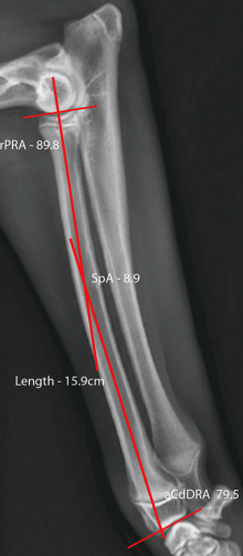
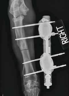
After a month of distraction, we can see the amount of bone length that was achieved (figure below). There is also a cone of new bone at each end of the osteotomy. When performing these distraction procedures, there is a fine balance in timing. If performed too slowly, the bone may heal before distraction is complete. If performed too quickly there is not enough time to allow the soft tissues (tendons, muscles and ligaments) to stretch with the bone, which may impede full extension of the carpus. During his distraction, Lito has also been involved in a rigorous formal physical rehabilitation program.
 |
Lateral radiograph after one month of distraction. Note the length of the distraction gap and the new bone forming from each fragment end.
Once Lito’s distraction program was completed and the desired radial length was achieved, the bone was permitted to consolidate. When complete healing occurred, his fixator was removed. He continues physical rehabilitation to improve his joint range of motion, build muscle mass and improve overall limb use.
 |
 |
Above: Lito’s radius has almost completely healed.